Tooth Decay & Cavities — Expert Treatment in Bangalore
Tooth decay is the most common dental disease worldwide, yet it is also the most preventable. It is not an inevitable condition. It doesn’t have to lead to pain, infection, or tooth loss. Additionally, it doesn’t always require drilling for treatment.
At Dental Solutions Clinic in Indiranagar, Bangalore, tooth decay is diagnosed and treated by Dr. Ramya Balasubramanya — a specialist Prosthodontist with MDS (Prosthodontics), BDS Gold Medallist — RGUHS University Topper, a Certified Digital Smile Design (DSD) Practitioner, and a Certified Invisalign Provider. Over more than 25 years of specialist practice, Dr. Ramya handles all restorative treatment at this clinic — from early-stage fillings and laser cavity preparation to crowns, inlays, full mouth rehabilitation, and laser-assisted root canal treatment.

What Is Tooth Decay?
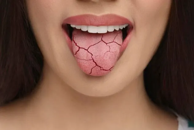
Tooth decay, also known as dental caries or cavities, is an infectious and transmissible disease caused by acid-producing bacteria residing in the mouth. It is more than just consuming sugar; it involves bacteria metabolising fermentable carbohydrates to produce lactic acid, which gradually demineralises the tooth structure. If untreated, decay progresses from the outer enamel through the softer dentine and eventually reaches the dental pulp — the nerve and blood supply at the tooth’s core.
This condition is the most common chronic disease worldwide, affecting more people than any other ailment. Fortunately, it is also highly preventable. Typically, the difference between someone who loses teeth to decay and someone who doesn’t is genetic, but it also depends on factors such as diet, oral hygiene practices, saliva quality, and early detection and treatment of initial lesions before they develop into cavities.
Symptoms of Tooth Decay
The most important clinical fact about tooth decay is that it is painless in its early and most treatable stages. Symptoms appear only when the disease has already progressed significantly:

No symptoms
Stage 1 and early Stage 2 decay produce no pain, no sensitivity, and no visible change that a patient would notice. Detection requires clinical examination and radiographs.

White or brown spot on the tooth surface
The earliest visible sign, a chalky or discoloured area on the enamel indicating subsurface demineralisation. Still reversible at this stage.

Sensitivity to sweet foods and drinks
Indicates the decay has reached the dentine layer, which contains tubules that transmit sensation to the nerve.

Sensitivity to cold or hot that lingers
More advanced dentine involvement; the pulp is beginning to respond to thermal stimuli.

Visible cavity or dark discolouration
The enamel has broken down, and a physical hole is present in the tooth.

Spontaneous toothache
A throbbing, continuous pain that occurs without any trigger indicates pulp involvement (pulpitis). This is a dental urgency.

Pain on biting
Can indicate a cracked tooth, abscess, or periapical infection associated with an untreated cavity.

Swelling, bad taste, or pus at the gum margin
Signs of a periapical abscess from a tooth with an infected or dead pulp. Requires immediate treatment.
Common Causes and Risk Factors

Frequent Sugar Consumption
The frequency of sugar exposure is more significant than the total amount. Sipping a sugary drink over two hours causes a prolonged acid attack, while drinking it in just 10 minutes results in a short, manageable challenge. The most common dietary risk factors for decay include frequent snacking, sugary tea or coffee consumed throughout the day, and acidic drinks like carbonated beverages and fruit juice.

Poor Oral Hygiene
Inadequate plaque removal, especially at contact points between teeth and along the gum margin, enables bacterial buildup and continuous acid production on the tooth surface. Electric toothbrushes generally eliminate more plaque than manual brushing for most individuals. Interdental cleaning methods, such as floss or interdental brushes, target plaque in areas inaccessible to regular toothbrushes.
Dry Mouth (Xerostomia)
Saliva serves as the mouth’s natural defence against decay by buffering acids, remineralising early lesions, and mechanically removing food debris from teeth. Conditions or medications that drastically reduce salivary production can increase the risk of rapid, extensive decay. Typical causes include antihistamines, antidepressants, antihypertensive drugs, and autoimmune diseases like Sjogren’s syndrome.
Deep Pits and Fissures
The biting surfaces of molars contain deep grooves, known as pits and fissures, which are challenging to clean with a toothbrush. These areas are the most frequent sites for early decay, especially in children and teenagers. Applying dental sealants, a thin protective coating to these surfaces, can effectively prevent fissure decay.
Stages of Tooth Decay
Initial Lesion (White Spot)
The earliest visible sign of decay is a chalky white or brown spot on the enamel surface. No cavity has formed. At this stage, the lesion can be arrested and reversed with fluoride application, dietary modification, and improved oral hygiene. No drilling required.
Enamel Decay
The lesion has extended into the enamel, creating a small cavity, but the dentine remains unaffected. The treatment is minimally invasive, involving the removal of decayed tissue and the placement of a small composite filling.
Dentine Decay
Decay has penetrated the dentine. Dentine decay progresses faster than enamel decay. Patients may experience sensitivity to sweet, cold, or hot stimuli. Treatment often requires a larger filling or, depending on the extent, an inlay, onlay, or dental crown.
Pulp Involvement
Bacteria have infiltrated the dental pulp, causing pulpitis. This condition results in a spontaneous toothache and, if untreated, leads to pulp death. The treatment typically involves root canal therapy, followed by placing a crown.
Abscess and Tooth Loss
An untreated pulp infection can spread to the adjacent bone, leading to a periapical abscess. Treatment involves either performing a root canal or extracting the tooth, then managing the bone and considering tooth replacement options.
How We Diagnose Tooth Decay
- Visual and tactile examination:
Identification of cavitations, enamel discolouration, and soft or sticky tooth surfaces - Digital radiography (bitewing X-rays):
Detection of interproximal (between-tooth) decay invisible to clinical examination - DIAGNOdent laser caries detection:
Quantifies mineral loss in fissures and smooth surfaces, detecting early lesions before they are visible - CBCT scan:
Where complex lesions, resorption, or extensive decay require three-dimensional assessment
How We Treat Tooth Decay

For Initial Lesions: Remineralisation and Prevention
Early white-spot lesions can be halted and reversed through the use of high-concentration fluoride, CPP-ACP products, dietary counselling, and better oral hygiene. No preparation or filling is necessary.

For Enamel and Early Dentine Decay: Minimally Invasive Fillings
Small to medium cavities are filled with tooth-coloured composite resin. Instead of a dental drill, Er:YAG laser preparation can be used to precisely remove decayed tissue without vibration, heat, or noise, often eliminating the need for local anaesthesia in the early stages.

For Deep Dentine Decay: Crown Restoration
Teeth with severe dentine decay that weaken the cusps need a full-coverage crown. Using our intraoral scanner for digital impressions and CAD/CAM technology, crowns can be designed and sometimes placed in just one visit.

For Pulp Involvement: Root Canal Treatment
Where bacteria have reached the dental pulp, root canal treatment removes the infected tissue, disinfects the canal system, and seals the tooth for long-term preservation. Laser-assisted root canal treatment using the Fotona LightWalker Er:YAG — performed by Dr. Ramya Balasubramanya — significantly improves disinfection of the canal system. Dr. Ramya also performs microscopic endodontics for complex, retreatment, and multi-rooted cases.
Frequently Asked Questions
Can a cavity heal on its own?
Very early lesions that haven’t yet cavitated can be remineralised through fluoride application and dietary modifications. However, once a cavity forms physically, it cannot heal on its own. The decay will persist until the bacterial infection is eliminated and the cavity is repaired.
Is a filling painful?
Fillings done with local anaesthesia are painless. For small enamel cavities, Er:YAG laser procedures usually don’t need anaesthesia. Sensitivity after filling in deeper lesions is common for 1–2 weeks but typically improves on its own.
How long do fillings last?
Composite resin fillings generally last between 7 and 12 years. Ceramic inlays and onlays can last 15 to 20 years or more. Amalgam fillings are rarely used at Dental Solutions Clinic now because composite and ceramic options offer similar or better durability and improved appearance.
My child has cavities in baby teeth. Does it matter?
Certainly. Untreated decay in primary teeth can cause pain and infection and may affect the developing adult tooth beneath. Baby teeth help preserve space for permanent teeth. Thus, decay in primary teeth should be addressed rather than neglected, believing they will eventually fall out.
Can dental sealants prevent tooth decay entirely?
Sealants are very effective in preventing pit and fissure decay on the biting surfaces of molar teeth, which are the most common areas for decay in children and teenagers. Research indicates that correctly applied sealants can decrease fissure decay by as much as 80%. However, they do not protect against decay between teeth or on smooth surfaces, and regular monitoring is necessary to keep them intact. Sealants are most advantageous when applied soon after the permanent molars erupt.